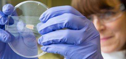
Part of the Faculty of Infectious and Tropical Diseases, and based at our Keppel Street site. The Imaging and Cytometry Platform for Infection Biology has been established to enhance the quality of research from staff at the School by providing access to, and specialised training for a range of equipment in pursuit of excellence within laboratory-based research. The equipment within this facility are open access to both staff and students of the School as well as external scientists. Please contact us to discuss your requirements.
Our staff
- Theresa Ward, Academic in Charge
- Liz McCarthy, Microscopy
- Christopher Chiu, Cytometry
Academic Focus Group: Theresa Ward, Rob Moon, Serge Mostowy, Francisco Olmo, Helena Helmby, Christiaan van Ooij, Amanda Fortes-Francisco, Fatoumatta Darboe, Archie Khan and Dominik Brokatzky.
Our Microscopy equipment
Zeiss LSM880 with Airyscan
- Location Keppel Street, lab 326
- This system has two detection modes, the conventional scanning confocal and the Airyscan which provides 1.7 fold improved resolution 8x improved signal to noise ratio. It is equipped with an environmental chamber for live cell imaging experiments.
- Lasers:
- 405nm laser diode
- 458, 488, 514 argon laser
- 561nm HeNe laser
- 594nm HeNe laser
- 633nm HeNe laser
Nikon Ti 2
- Location Keppel Street, lab 333B
- Equipped with OKO-lab environmental controls (CO2 and Nitrogen) and fully motorized stage allowing for live cell imaging, tile scanning and the imaging of multiple locations. The piezo stage provides high speed z-axis control, perfect for rapid z-stack acquisition. The system comes equipped with the Perfect Focus System (PFS) for focus stabilisation of live cell imaging. A variety of stage inserts and objectives (including extra long working distance, ELWD, lenses) enables samples in a variety of formats including standard slides, dishes and multi-well plates to be imaged.
- LED lines:
- 365, 385, 405, 435
- 460, 470, 490, 500
- 525, 550, 580, 595
- 635, 660, 740, 770
Nikon Ti E _ Fixed cell
- Location Keppel Street, lab 326
- The sister microscope to the Nikon in 333B has a variety of stage inserts and objectives enables samples in a variety of formats including standard slides, dishes and multi-well plates to be imaged.
- Fluorescence illumination source – mercury bulb.
Image Analysis
Located in office 490 at Keppel Street, we have a dedicated high –end image analysis computer. In addition to the open source softwares: Image J/FIJI and Cell Profiler. Staff and students of the School can use Volocity from PerkinElmer, or NIS-Elements from Nikon as well as Zen from Zeiss. Guidance and/or consultations for your specific requirements are available.
Contact person:
Dr Liz McCarthy, Faculty Microscopy Manager
E: elizabeth.mccarthy@lshtm.ac.uk
T: +44 (0)20 7927 2063
Our Flow Cytometry equipment
Cytek Aurora
- Location: Keppel Street, lab 346
- Description: The Cytek Aurora is a 4 laser spectral cytometer which allows for:
- Measurement of up to 35 colours from one sample
- High sample throughput with a plate/tube loader.
- Small particle detection, to look at particles down to approximately 70nm
- Lasers:
- Violet 405nm
- Blue 488nm
- Yellow Green 561nm
- Red 640nm
- Restrictions: Fixed samples only
Thermo Fisher Scientific Attune NxT flow cytometry cell analyser
- Location: Keppel Street, lab 382
- Description: This is a 4-laser system allowing for analysis of up to 14 fluorescent parameters, with an integrated autosampler for acquiring samples from 96-well and 384-well plates
- Lasers:
- Violet 405nm
- Blue 488nm
- Yellow 561nm
- Red 640nm
- Restrictions: Housed within the Malaria containment level 3 suite
Becton Dickinson FACSAria™ Fusion flow cytometry cell sorter
- Location: Keppel Street, lab 426
- Description: This is a 3-laser system allowing for analysis of up to 10 fluorescent parameters
- Lasers:
- Blue 488nm
- Yellow 561nm
- Red 633nm
- Restrictions: Housed in a Class II Baker Biosafety cabinet within the main Aerosol containment level 3 suite
- Nozzle sizes:
- 70µm
- 85µm
- 100µm
- Sorting options:
- 4-way tube
- 6, 24, 48, 96 and 348-well plates
Data Analysis
An analysis workstation with a FlowJo software licence is available in Keppel street.
A USB dongle with a FlowJo software licence is available for remote access.
Contact person:
Christopher Chiu, Faculty Flow Cytometry Manager
T: +44 (0)7927 589935
Other equipment
If the technology you’re interested in is not listed here, please contact us to have a chat. We have good connections both internally and externally to establish collaborative work. Individual research groups at the School have imaging capabilities from histology sectioning and imaging equipment through to an in vivo IVIS imaging system.
